Amyloid-? (1-8, A2V) Peptide
Amyloid-? (1-8, A2V) Peptide
Amyloid-? (1-8, A2V) is a truncated form of amyloid-? (A?) that contains a valine to alanine substitution at position 2...
€114.00
View Rabbit Monoclonal Antibody")
€672.00 €571.20
Programmed cell death protein 1 (PD-1), also known as CD279, is a cell surface receptor belonging to the immunoglobulin superfamily that is involved in regulation and attenuation of the adaptive immune response.{52533,53686} It is a 288-amino acid type I transmembrane protein encoded by the PDCD1 gene in humans and is comprised of a 167-amino acid ectodomain consisting of an N-loop, IgV-like domain, and a stalk region, a transmembrane domain, and a cytoplasmic tail with two tyrosine-based signaling motifs.{53687,53689,53686,52533} PD-1 is expressed in activated immune cells including CD4+ T cells, CD8+ T cells, natural killer T (NKT) cells, B cells, monocytes, and dendritic cells.{53690,53686} Binding of PD-1 to either of its ligands, PD-L1 (Item No. 28378) or PD-L2 (Item No. 28379), suppresses T cell proliferation and cytokine production.{52533} PD-1 deficiency induces cardiomyopathy or lupus-like glomerulonephritis in BALB/c and C57Bl/6 mice, respectively.{53687,53686} Increased levels of intratumoral PD-1+ immune cells are associated with increased tumor size, higher nuclear grade, and poor prognosis in patients with renal cell carcinoma.{53688} Formulations containing PD-1 blocking antibodies have been used in the treatment of various cancers. Cayman’s PD-1/CD279 (N-Term) Rabbit Monoclonal Antibody can be used for immunohistochemistry (IHC) and Western blot (WB) applications.
Territorial Availability: Available through Bertin Technologies only in France
| Size | 100 µl |
|---|---|
| Shipping | dry ice |
| Molecular weight | 0 |
| Host | Rabbit |
| Antigen | Peptide from the N-terminal region of human PD-1 |
| Clone | RM309 |
| Isotype | IgG |
| Application(s) | IHC, WB |
| Formulation | 100 µl of protein A-affinity purified monoclonal antibody |
| Custom code | 3822.19 |
| UNSPSC code | 12352203 |
Cayman Chemical’s mission is to help make research possible by supplying scientists worldwide with the basic research tools necessary for advancing human and animal health. Our utmost commitment to healthcare researchers is to offer the highest quality products with an affordable pricing policy.
Our scientists are experts in the synthesis, purification, and characterization of biochemicals ranging from small drug-like heterocycles to complex biolipids, fatty acids, and many others. We are also highly skilled in all aspects of assay and antibody development, protein expression, crystallization, and structure determination.
Over the past thirty years, Cayman developed a deep knowledge base in lipid biochemistry, including research involving the arachidonic acid cascade, inositol phosphates, and cannabinoids. This knowledge enabled the production of reagents of exceptional quality for cancer, oxidative injury, epigenetics, neuroscience, inflammation, metabolism, and many additional lines of research.
Our organic and analytical chemists specialize in the rapid development of manufacturing processes and analytical methods to carry out clinical and commercial GMP-API production. Pre-clinical drug discovery efforts are currently underway in the areas of bone restoration and repair, muscular dystrophy, oncology, and inflammation. A separate group of Ph.D.-level scientists are dedicated to offering Hit-to-Lead Discovery and Profiling Services for epigenetic targets. Our knowledgeable chemists can be contracted to perform complete sample analysis for analytes measured by the majority of our assays. We also offer a wide range of analytical services using LC-MS/MS, HPLC, GC, and many other techniques.
Accreditations
ISO/IEC 17025:2005
ISO Guide 34:2009
Cayman is a leader in the field of emerging drugs of abuse, providing high-purity Schedule I-V Controlled Substances to federally-licensed laboratories and qualified academic research institutions for forensic analyses. We are certified by ACLASS Accreditation Services with dual accreditation to ISO/IEC 17025:2005 and ISO Guide 34:2009.
Amyloid-? (1-8, A2V) is a truncated form of amyloid-? (A?) that contains a valine to alanine substitution at position 2...
Protein phosphorylation is an important post-translational modification that serves many key functions to regulate a protein’s activity, localization, and protein-protein...
To be used in conjunction with Cayman’s FABP3 polyclonal antibody (Catalog No. 10233) to block protein-antibody complex formation during immunochemical...
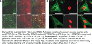
Prostaglandin I synthase (PGIS) catalyzes the isomerization of PGH2 to PGI2. PGI2 (prostacyclin) is a potent vasodilator and inhibitor of...
To be used in conjunction with Cayman’s NAPE-PLD polyclonal antibody (aa 6-20) (Item No. 10306) to block protein-antibody complex formation...
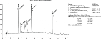
This mixture contains the characteristic metabolites of both PGI2 and TXA2. Contents: Thromboxane B2, 11-dehydro Thromboxane B2, 6-keto Prostaglandin F1?,...
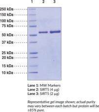
Sirtuin 5 (SIRT5) is an enzyme that catalyzes the NAD-dependent removal of malonyl, succinyl, and glutaryl groups from target proteins.{55107,55108}...
This mixture contains the primary COX products produced by most mammalian tissues. Contents: Prostaglandin D2, Prostaglandin E2, 6-keto Prostaglandin F1?,...
TAF10 is one of many protein factors or coactivators associated with RNA polymerase II activity.{16903} One vial of this peptide...
The cyclopentenone prostaglandin HPLC mixture contains all of the major UV-absorbing cyclopentenone prostaglandins and their precursors supplied in methyl acetate....
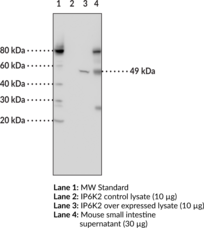
Inositol hexakisphosphate kinase 2 (IP6K2) is a cytoplasmic kinase that catalyzes the conversion of IP6 to diphosphoinositol pentakisphosphate in the...

Platelet-activating factor (PAF) is an important lipid mediator involved in inflammation. PAF-acetylhydrolase (PAF-AH) is an extracellular phospholipase A2 which hydrolyzes...
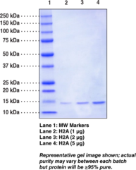
Histone H2A is a core histone that forms a dimer with histone H2B.{46631} Two histone H2A/H2B dimers form an octameric...
The Cayman COX Inhibitor Pack contains a combination of frequently used cyclooxygenase (COX) inhibitors. Each kit contains aspirin, the archetype...
GST-tag Polyclonal Antibody (FITC) is a probe for the immunochemical detection of GST tags on recombinant proteins. Recombinant proteins are...
This mixture contains primary prostaglandins produced from arachidonic acid and dihomo-?-linolenic acid. Contents: Prostaglandin E1, Prostaglandin E2, Prostaglandin F1?, 6-keto...
Programmed cell death protein 4 (PDCD4) levels are elevated during apoptosis and absent in many cancer samples.{16340,16167} Loss of PDCD4...
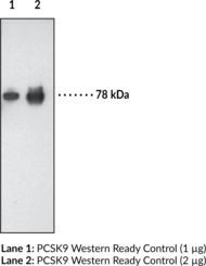
Proprotein convertase subtilisin kexin 9 (PCSK9) is a member of the subtilisin serine protease family with an important role in...